Question 1#
A 71-year-old man with a history of hypertension and hyperlipidemia presents with severe left knee pain and fever. Physical examination demonstrates a swollen left knee with limited range of motion. The patient is noted to have a temperature of 38.8°C and a blood pressure of 150/100 mmHg. Synovial fluid is drawn from the left knee and reveals the following.
- Leukocyte count 28,000/mm3 (94% neutrophils)
- Crystals Positively birefringent, rhomboid-shaped
- Gram stain Negative
Which of the following is likely to be present in this patient’s condition?
A. Calcification of articular cartilageB. Rheumatoid factor
C. Tophi
D. S. aureus bacteremia
Correct Answer is A
Comment:
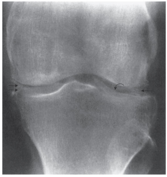
Calcification of articular cartilage. The patient in this question is showing signs and symptoms of pseudogout, an acute arthritis that is caused by calcium pyrophosphate dihydrate (CPPD) crystal release from calcified articular cartilage into the joint space. This calcification of articular cartilage is referred to as chondrocalcinosis and often manifests with pain, swelling, erythema, fever, and limited range of motion. The knee is the most common joint that is affected in pseudogout (Figure below demonstrates articular cartilage calcification on x-ray).
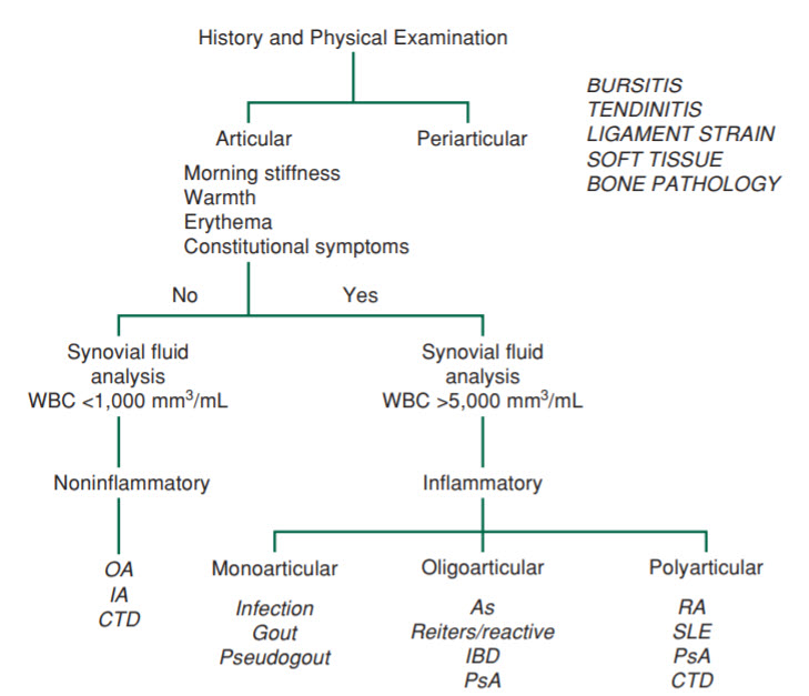
Of note, even though fever and leukocytosis (often with left shift as seen in this patient) can occur in pseudogout, they are not required for the diagnosis. Diagnosis of pseudogout relies on identifying rhomboidshaped positively birefringent crystals on synovial fluid analysis. An approach to the initial differential diagnosis of arthritis is shown below in Figure below.
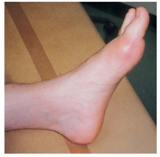
(B) Rheumatoid factor (RF) typically occurs in rheumatoid arthritis (RA); however, a positive RF does not make the diagnosis of RA as RF can be positive in several other conditions. RA is characterized by a symmetric arthritis of insidious onset. Fever can be present as well. (C) Tophi are characteristic of gout (specifically chronic gout) and are urate crystals that manifest as yellowish nodules of firm consistency at affected joints (Figure below).
Gout is diagnosed by needle-shaped, negatively birefringent crystals on synovial fluid analysis. (D) S. aureus bacteremia with arthritis would characterize septic arthritis (S. aureus is the most common cause). Even though this patient presents with fever and leukocytosis, the identification of rhomboid-shaped positively birefringent crystals leads to the diagnosis of pseudogout. Furthermore, the gram stain is negative with this patient.