Question 1#
A 22-year-old man with a history of severe reactive airway disease and polysubstance abuse is brought to the emergency department following a motor vehicle collision. Endotracheal intubation is performed and placement confirmed by continuous capnography. Peak inspiratory pressure on the ventilator is 45 cm H2O.
Which of the following describes the MOST LIKELY appearance of his capnogram?
A. Box shaped with a flat phase IIIB. Biphasic with a rounded phase III
C. Shark finned with up-sloped phase III
D. Flat
Correct Answer is C
Comment:
Correct Answer: C
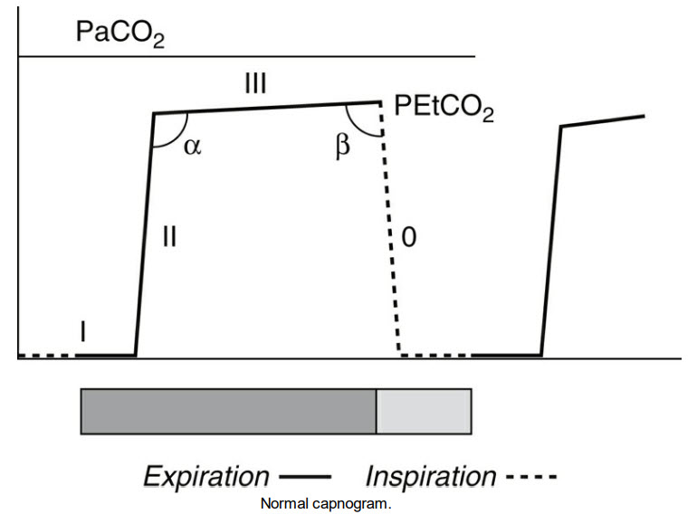
A timed capnograph has 4 segments with phase 0 representing inspiration. In a mechanically ventilated patient, a “normal” capnograph takes a relatively box-shaped appearance with a relatively flat phase III (A, Normal Capnogram figure).
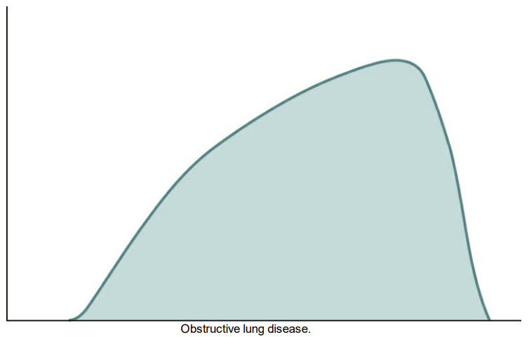
The clinical scenario in the question suggests obstructive lung pathology which manifests as an up-sloped phase III which some have described as “shark-finned” appearance (C, Obstructive Lung Disease figure). Any airway obstruction, including obstruction of the endotracheal tube, can result in this waveform. A biphasic capnograph has been previously described but is not specific for endobronchial intubation (B). Regardless, its presence should prompt further investigation.
Capnography has additional utility as a noninvasive cardiac output monitor. Current American Heart Association/ Advanced Cardiac Life Support guidelines recommend the use of continuous capnography during cardiopulmonary resuscitation as an assessment for adequacy of cardiopulmonary resuscitation and a sensitive detector for return of spontaneous circulation. A flat capnograph should alert the physician to possible esophageal intubation or inadvertent extubation, as adequate ventilation should produce a waveform (D).
References:
- Cook TM, Woodall N, Harper J, Benger J. on behalf of the Fourth National Audit Project. Major complications of airway management in the UK: results of the Fourth National Audit Project of the Royal College of Anaesthetists and the Difficult Airway Society. Part 2: intensive care and emergency departments. BJA Br J Anesth. 2011;106(5):632-642.
- Bhavani Shankar Kodali. Capnography outside the operating rooms. Anesthesiology. 2013;118(1):192-201.
- Link MS, Berkow LC, Kudenchuk PJ, et al. Part 7: adult advanced cardiovascular life support: 2015 American Heart Association Guidelines Update for Cardiopulmonary Resuscitation and Emergency Cardiovascular Care. Circulation. 2015;132(suppl 2):S444-S464.