Question 9#
A 5′7″ middle-aged man, with unknown medical history, has just arrived in the operating room for emergent exploratory laparotomy following intubation in the emergency department with appropriate hypnotic and 100 mg rocuronium. The initial ventilator settings are tidal volume 450 mL, rate 16 breaths/min, PEEP 5 cm H2O and FiO2 40%, and inspiratory:expiratory (I:E) ratio 1:2. The EtCO2 waveform follows.
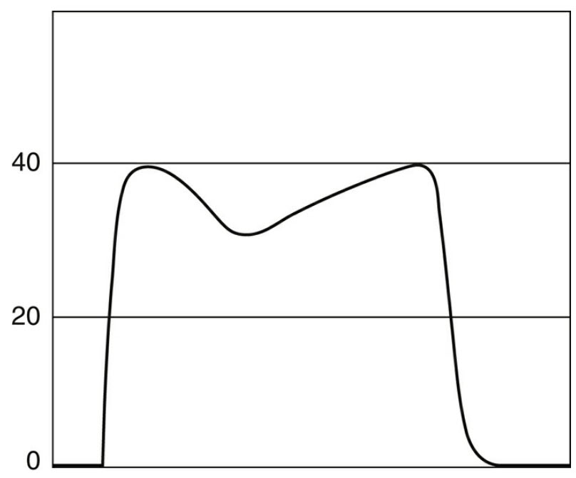
Based on the EtCO2 waveform, what would be the BEST next course of action?
A. Give an additional dose of muscle relaxationB. Increase minute ventilation to 600 mL
C. Continue current ventilator setting
D. Change the I:E ratio to 1:3
Correct Answer is D
Comment:
Correct Answer: D
A biphasic EtCO2 waveform suggests that the patient had undergone single lung transplantation, most likely from severe emphysema. The first peak shows CO2 emptying of an allograft with normal compliance and airway resistance. The second peak reflects the obstruction of the native emphysematous lung. Since the emphysematous lung is more compliant than the donor lung, most of the tidal volume will preferably go to the native lung. As a result, the native lung, which has a severe expiratory obstruction, is prone to hyperinflation and auto-PEEP. Therefore, expiratory time should be allowed as long as possible to allow time to completely empty the native lung (answer D).
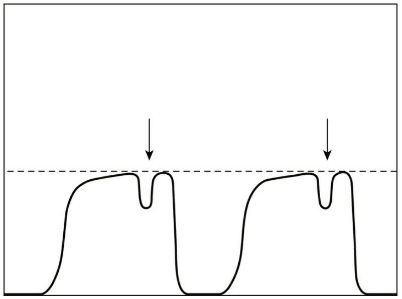
Answer A is incorrect because the waveform does not show recovery from muscle relaxation. When the patient recovers from muscle relaxation, a curare cleft will appear in the EtCO2 tracing as shown below:
Answer B is incorrect because the patient predicted body weight given his height and gender is around 66 kg. A tidal volume of 600 mL then would be 9 mL/kg IBW. Since we want to use lung protective ventilation, especially in a patient with a history of lung transplant, this tidal volume would be too high.
References:
- Single Lung Transplant. Available at https://aneskey.com/wpcontent/uploads/2017/06/image01049.jpeg.
- Capnography. Available at https://www.csems.org/wpcontent/uploads/2018/04/Capnography.png.
- Rai HS, Boehm JK, Stoller JK. Biphasic capnogram in a single-lung transplant recipient: a case report. Respir Care. 2014;59(8):E108-E109.
- Barnes L, Reed RM, Parekh KR, et al. Mechanical ventilation for the lung transplant recipient. Curr Pulmonol Rep. 2015;4(2): 88-96.